FIGURE 2.
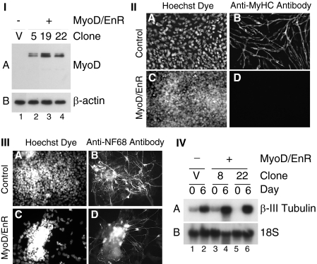
Overexpression of MyoD/EnR inhibited the formation of skeletal myocytes but not neurons in aggregated P19 cells. I, P19(Control) (lane 1) and P19(MyoD/EnR) cells (lanes 2–4) were grown in monolayers, and total protein was harvested. Western blots with 30 μg of total protein were probed with antibodies indicated on the right. II, P19(Control) cells (panels A and B) and P19(MyoD/EnR) cells (panels C and D) were differentiated in the presence of 0.8% DMSO. On day 9 of the differentiation, cells were fixed and stained with Hoechst dye to detect nuclei (panels A and C) and anti-MyHC antibody (panels B and D). III and IV, P19(Control) and P19(MyoD/EnR) cells were differentiated in the presence of 1 μm retinoic acid. III, on day 6 of the differentiation, cells were fixed and stained with Hoechst dye to detect nuclei (panels A and C) and anti-NF68 antibody (panels B and D). IV, Northern blots with 4 μg of RNA were probed with the factors indicated on the right. Lanes are indicated at the bottom of I and IV. Magnification ×400 for II and III.

