FIGURE 3.
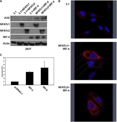
NFAT and IRF are necessary and sufficient for induction of A3G expression. A, 293T cells were co-transfected with the indicated expression plasmids. Whole cell lysates were probed for expression of transfected plasmids and A3G. Immunoblots probed for actin expression are also shown (loading control). B, 293T cells were transfected with the indicated plasmids, and cells were stained for A3G. Nuclei were counterstained with DAPI. C, 293T cells were transfected with pcDNA3.1, IRF-4, or IRF-8 plus reporter construct. Relative promoter activity was determined by comparing the IRF-4 and IRF-8 signal with the pcDNA3.1 signal.

