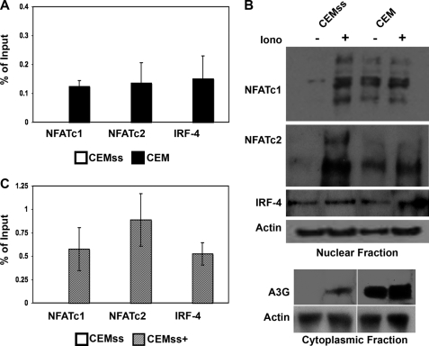
FIGURE 4.
Enrichment of nuclear NFAT induces transactivation of the A3G promoter in T cells. A, antibodies against NFATc1, NFATc2, IRF-4, and an Ig control were used in a ChIP assay. Immunoprecipitated samples from CEMss and CEM were analyzed in a TaqMan real-time PCR assay using primers directed to the A3G minimal promoter region. The average cycle threshold of GAPDH for input samples was used as an internal control for quality of input DNA, CEMss Ct 27.4 ± .87, CEM Ct 27.3 ± .78. B, Mock-treated (−) and ionomycin (Iono)-stimulated (+) CEMss and CEM cell lysates were subcellularly fractionated and probed for NFATc1, NFATc2, or IRF-4 expression in the nuclear fraction. A3G expression was assayed in the cytoplasmic fraction. Actin expression is shown as a loading control. C, CEMss and ionomycin stimulated CEMss (CEMss+) cells were assayed for NFATc1, NFATc2, or IRF-4 occupation of the A3G promoter as in A. Average cycle threshold of GAPDH for input samples was used as an internal control for quality of input DNA; CEMss Ct 27.4 ± .87, CEMss+ Ct 28.2 ± .78. Ct, threshold cycle.

