FIGURE 4.
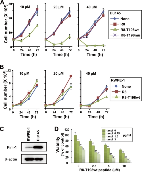
Growth inhibitory effect of cell-permeable p27Kip1 peptide. A, DU145 cells were treated with the indicated concentrations of FITC-labeled cell-permeable Arg8, R8-T198wt, or R8-T198mu peptide. Viable cells were counted after incubation for 24, 48, and 72 h. Each vertical bar represents the mean ± S.D. of three independent experiments. B, RWPE-1 cells were treated as in A. Viable cells were counted after incubation for 24, 48, and 72 h. Each bar represents the mean ± S.D. of three independent experiments. C, total cell lysates of RWPE-1 and DU145 cells were electrophoresed and immunoblotted with the indicated antibodies. D, DU145 cells were incubated in a medium containing the indicated concentrations of the FITC-labeled R8-T198wt peptide (0–10 μm) together with the indicated concentrations of taxol (0–3 μg/ml). Viable DU145 cells were counted by the trypan blue dye exclusion method. The number of viable untreated control cells was normalized to 100%. Each vertical bar represents the mean ± S.D. of three independent experiments. The interaction of various concentrations of the R8-T198wt peptide and taxol was subjected to two-way analysis of variance. *, p < 0.05; **, p < 0.01.

