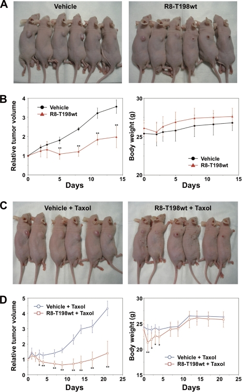
FIGURE 5.
In vivo anti-tumor activity of p27Kip1 peptide. A and B, DU145 cells were subcutaneously implanted into the right flank of BALB/c nude mice. Therapeutic experiments (six mice/group) were started (day 0) when the tumor reached a volume of ∼100 mm3. The R8-T198wt peptide was administered by intratumoral injection in 50 μl of 10 mm solution on days 0 and 3. The control group received the same volume of vehicle on days 0 and 3. A, representative surface tumor morphology of the mice on day 8 after initiation of treatment (left, vehicle; right, R8-T198wt peptide). B, relative tumor volume and body weight. C and D, R8-T198wt peptide (50 μl of 10 mm) or the same volume of vehicle was administered intratumorally on days 0 and 5. All DU145-bearing mice were intravenously administered 60 mg/kg taxol on day 0. C, representative surface tumor morphology on day 14 after initiation of treatment (left, vehicle and taxol; right, R8-T198wt peptide and taxol). D, relative tumor volume and body weight. *, p < 0.05; **, p < 0.01.

