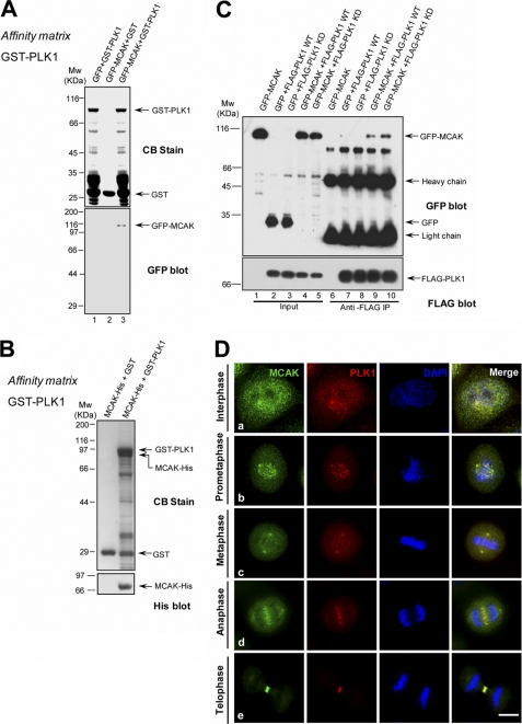
FIGURE 1.
MCAK interacts with PLK1 in vitro and in vivo. A, GST-PLK1 on glutathione beads was incubated with lysates of 293T cells transiently transfected with GFP or GFP-MCAK. The beads were washed and analyzed by Western blotting with an anti-GFP antibody. CB, Coomassie Blue stain. B, GST-PLK1, but not GST, pulled down MCAK-His. GST-PLK1 on glutathione-conjugated beads was incubated with recombinant MCAK-His protein purified from Sf9 cells, followed by Western blot analysis using an anti-His antibody. C, co-immunoprecipitation of FLAG-PLK1 and GFP-MCAK. 293T cells were transiently transfected with GFP-MCAK plus FLAG-PLK1 WT (wild type) or FLAG-PLK1 kinase-dead (K82A). 36 h post-transfection, cells were lysed and incubated with the anti-FLAG antibody coupled to agarose beads. The beads were washed and analyzed by Western blotting with an anti-GFP mouse antibody and then probed with an anti-FLAG antibody. D, co-localization of endogenous MCAK and PLK1 during mitosis. HeLa cells were synchronized to mitosis by a thymidine block release, fixed, and then stained with an anti-MCAK rabbit antibody and an anti-PLK1 mouse antibody. DNA was stained with DAPI. MCAK is labeled in green and PLK1 is in red. Scale bar, 10 μm.

