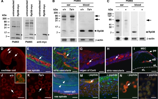
FIGURE 6.
Tissue expression and immunolocalization of Rpl38. A–C, Western blots of protein extracts from HEK293T cells (A) and tissue extracts of adult +/+ and Ts/+ mice (B) in the presence (+) and absence (−) of Pb863-specific peptide (C). Left side of each panel indicates the molecular mass in kDa. A, membranes stained with Pb863, Pb865, and anti-Myc antibody of HEK293T cells transfected and nontransfected with a mouse RPL38-myc fusion construct. Black dot indicates Rpl38-specific band present in the transfected but not in untransfected lysate. B, Western blot of protein extract from the ear and blood of +/+ and Ts/+ mice blotted with Pb865 and β-actin antibody. Arrowhead indicates the ∼8-kDa Rpl38 protein band. Arrows point to higher molecular weight bands in ear lysate. Protein lysates were also hybridized with β-actin antibody to control for equal loading. C, ear and blood protein lysates were incubated with Pb865 in the presence (+) and (−) absence of Pb865-specific peptide. Note that both the ∼8-kDa and higher molecular weight protein species are effectively blocked by the peptide. Membranes were hybridized with β-actin antibody to control for equal transfer efficiency. D--N, shown are confocal images of cochlear coil (D and E) and stria vascularis (F) whole mount preparations, cryosections of organ of Corti (G), stria vascularis (H), otic capsule (I), and blood spreads (J and K) stained with the Pb865 antibody (red channel). E–I and L--N, counterstained with DAPI (blue channel); E and F, counterstained with phalloidin (green channel); L, staining with nonspecific rabbit IgG antibody. J, inset shows high magnification of Z-stack of Rpl38-positive erythrocyte showing staining near the plasma membrane. White arrows (D–N) point to Rpl38-positive staining, and arrowhead in L points to nonstaining erythrocytes. M and N, staining of stria vascularis (M) and blood spread (N) with Pb865 in the absence (−) and presence (+) of Pb865-specific peptide. Note that Pb865 staining is blocked in the presence of competing peptide. pe, periosteum; eb, endochondral bone; el, endosteal layer; sl, spiral ligament; MEC, middle ear cavity. Scale bars, E and F, 10 μm; G and H, 20 μm; I, 50 μm; J, K and N, 5 μm; L and M, 20 μm.

