FIGURE 7.
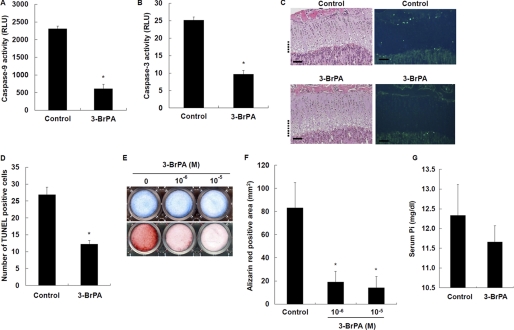
Effects of 3-BrPA on chondrocyte apoptosis and calcification. A, effects of 3-BrPA on caspase-9 activity. B, effects of 3-BrPA on caspase-3 activity. Cells were treated with 10−6 m 3-BrPA for 7 days and measured for caspase activity. C, histological examination of chondrocyte apoptosis. Hematoxylin/eosin staining (left) and TUNEL staining (right) were performed on tibial sections from 31-day-old control and 3-BrPA-treated mice. The hypertrophic zone is marked with dotted lines, and the scale bars indicate 200 μm. D, number of TUNEL-positive cells in tibial growth plates of control and 3-BrPA-treated mice. E, histochemical staining of chondrocytes. Cells were cultured for 7 days in the presence of 10−6 and 10−5 m 3-BrPA, and stained with Alcian blue for GAG synthesis (upper) and alizarin red S for mineralization (lower). F, quantification of alizarin red staining. G, serum Pi levels. Results are expressed as the mean ± S.E. of four separate experiments. *, significantly different from control (p < 0.05). RLU, relative light units.

