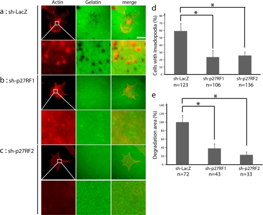
FIGURE 4.
p27RF-Rho regulates gelatin degradation activity in F10 cells. a, degradation of Oregon Green-labeled gelatin (middle column) and actin immunohistochemistry (red, left column) in control (sh-LacZ) and p27RF-Rho-depleted (sh-p27RF1 and sh-p27RF2) cells cultured on glass coverslips (scale bar, 20 μm). Higher magnification views of the boxed areas are shown underneath each image. b, quantification of cells with invadopodia. The cells were scored as invadopodia-positive if they contained punctate structures positive for both actin filaments and gelatin degradation. c, quantification of gelatin degradation area. The data are presented as the means ± standard errors of means (n = 5). The number of cells analyzed in each data set is indicated at the bottom of the data bars in the graphs. *, p < 0.05.

