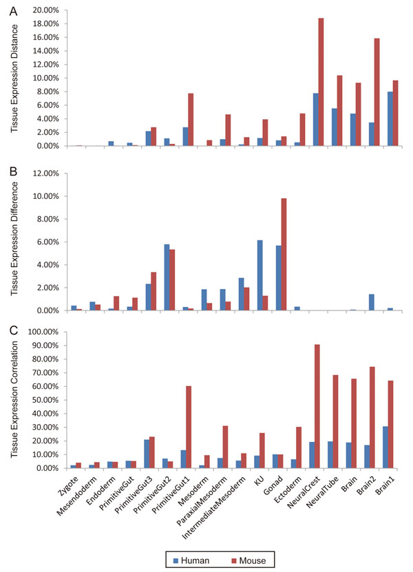
Figure 3.
Comparisons of conservatively expressed GO modules (A), differentially expressed GO modules (B), and correlatively expressed GO modules (C) between human and mouse tissues. Red bars denote the mouse tissues, and blue bars the human tissues. Each bar height represents the percentage of significant GO modules (p<0.05) out of the total number of GO modules analyzed from mouse or human. The names of tissue group nodes on the X axis are consistent with those used in Table 1.

