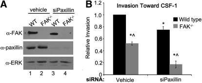
Figure 3. FAK and paxillin regulate CSF-1-dependent macrophage invasion through separate signaling pathways.
(A) Immunoblot analysis showing paxillin knockdown under control (Lanes 1 and 2) and siPaxillin-treated conditions (lanes 3 and 4) in WT and FAK−/− BMMs. (B) Relative invasion toward CSF-1 of WT and FAK−/− BMMs treated with vehicle or siRNAs targeting paxillin. *Values that are significantly different from vehicle-treated WT cells; ^values that are significantly different from WT cells under the same treatment conditions; n = 6.

