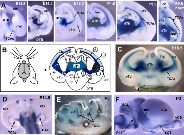
Figure 1.
The TCA-TLZ reporter line marks thalamocortical axons specifically and consistently during development. (A) The TCA-TLZ reporter expresses beta-galactosidase in dorsal thalamic neurons (dTh) starting from E13, and reveals the development of their axon trajectory (TCAs) to cortex (ctx). Cortical axons are not labeled by the reporter. Olfactory axons are labeled in the anterior commissure (aca); some cells in hippocampus (hip) label postnatally. Coronal vibratome sections (100 μm) of brains of indicated ages were stained with X-Gal. The postnatal (P)9.5 sample is cut horizontally to show TCAs fanning out. ob, olfactory bulb. (B) Schematic of TCA pathway as viewed in a coronal section of a P0 mouse brain, with developmental steps numbered. See text for details. TCAs 1) grow ventrally; 2) turn to cross the diencephalon-telencephalon border (DTB) by E13.5; 3) defasciculate and fan out in striatum (Str); 4) cross the corticostriatal boundary (CSB) and turn dorsally into cortex; 5) extend dorsally in a restricted layer; 6) make collateral branches into the cortical target area. Hy, hypothalamus; i.c., internal capsule; LV, ventricle. (C) The cut face of the caudal half of an E18.5 brain expressing the TCA-TLZ transgene shows the TCA projection from the dorsal thalamus through the ventral telencephalon (vTel) and into cortex. The hippocampus (hip) fills the lateral ventricle. The optic tract (ot) is also labeled by the reporter. (D) Dorsal view of a whole-mount E14.5 brain stained with X-Gal reveals the TCAs in the internal capsule (arrows). (E) A whole newborn TCA-TLZ brain was cut coronally in half and stained with X-Gal allowing visualization of TCA pathfinding in a whole brain. mb, midbrain. (F) A lateral view of a newborn TCA-TLZ brain stained with X-Gal shows labeling in the lateral olfactory tract (LOT) from the accessory olfactory bulb (AOB) and the pontocerebellar tract (PCT). TCAs beneath cortex produce light blue staining. Dark blue staining in entorhinal cortex (ent) is due to cellular staining in a superficial layer; TCAs do not project to entorhinal cortex. EGL, external granular layer of cerebellum.

