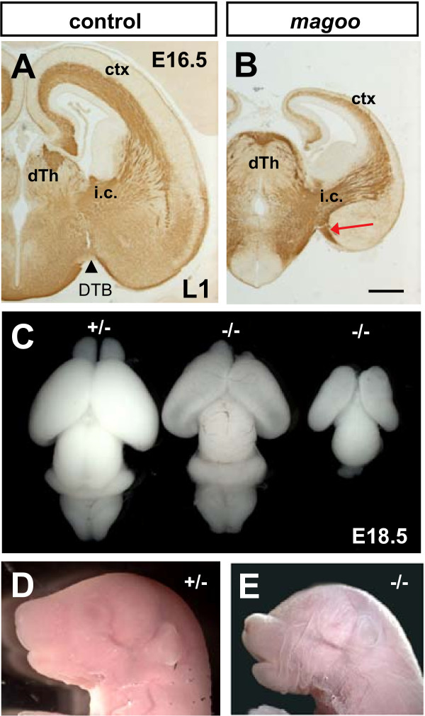
Figure 4.
magoo mutants have small malformed brains and craniofacial defects. (A, B) L1 immunolabels TCAs and corticothalamic axons in E16.5 brains. The approximate position of the DTB is indicated by a black arrowhead. In the magoo mutant brain, an abnormal axon bundle is seen extending ventrally off the internal capsule (i.c.) in the vTel, adjacent to the DTB (red arrow). ctx, cortex. Scale bar, 0.5 mm. (C) A heterozygote brain, left, with normal size and morphology was photographed next to two homozygous magoo mutant brains from the same E18.5 litter. The homozygote in the center has a slightly smaller brain with hollow lateral ventricles, and its right olfactory bulb is smaller than the left, not damaged. The homozygote brain at right is very small with no olfactory bulbs. (D) A normal E18.5 mouse head. (E) A homozygous magoo mutant E18.5 with small head, shortened snout, and microphthalmia.

