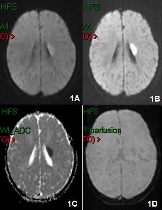
Figure 1.

Brain MRI with Gd-DTPA was performed and showed the presence of an hyperacute ischemic lesion of about 2 cm of diameter, in the left lenticular nucleus extended to the internal capsule 1A), 1B), 1C) in DWI and 1D) perfusion weighted sequences.
