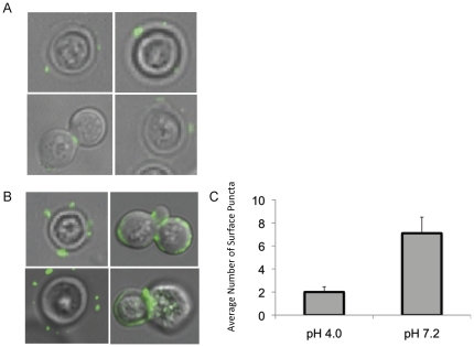
Figure 2. Localization of GlcCer in Cn grown in vitro at high CO2 and either acidic or neutral pH.
Indirect immunofluorescence was used to determine the localization of GlcCer in wild type Cn in media of either acidic (A) or neutral (B) pH. Primary antibody used is an anti-Cn GlcCer monoclonal antibody developed by our lab. The secondary was an isotype-specific FITC-conjugated antibody, and confocal microscopy was used to analyze the images. The amount of surface puncta per cell was quantified by counting puncta from three large fields of cells, averaged by cell number (C).

