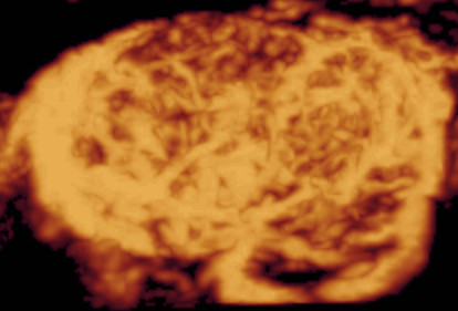
Figure 1.

Three-dimensional power Doppler ultrasound of Hürthle cell adenoma: presentation of the whole nodule. On the 3D rendered image of the whole nodule the peripheral and central vessels overlap making the evaluation of the abundant vascularization difficult. (See also: Additional file 1).
