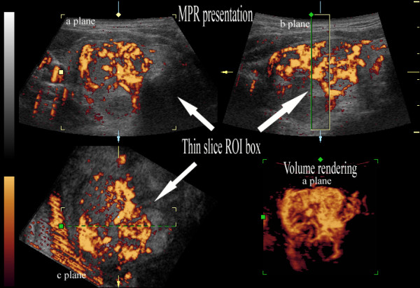
Figure 5.

Three-dimensional power Doppler ultrasound of papillary cancer with high density of vessels. Multiplanar reformation (MPR) mode presentation and thin-slice volume rendering (lower right corner). The density of the vessels is in the highest range: 4 = (76-100%) of area of the nodule; ROI - region of interest.
