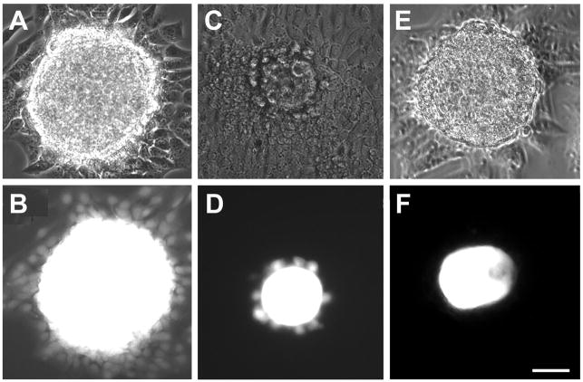
Fig. 5.
Gap junctions couple oocytes and granulosa cells into a functional syncytium. A, C, and E are phase contrast micrographs of cultured oocyte-granulosa cell complexes while B, D, and F are the corresponding fluorescence micrographs. (A,B) The existence of gap junctional coupling is revealed by spreading of fluorescent dye (Lucifer yellow), injected into the oocyte, throughout the surrounding granulosa cell population of a wildtype complex. (C,D) In CX43 null mutant follicles, dye injected into the oocyte is restricted from spreading beyond the first layer of granulosa cells due to the absence of gap junctions in the granulosa cells. (E,F) In CX37 null mutant follicles, the injected dye fails to pass into the granulosa cells owing to the absence of gap junctions linking them with the oocyte. Scale bars, 50 μm. Micrographs from Veitch et al. (2004) and Li et al. (2007) are republished with permission from the Company of Biologists Ltd.

