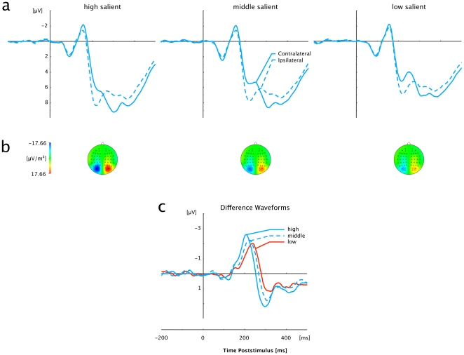
Figure 4. Grand averaged event-related brain potentials elicited in response to orientation-defined (pop-out) targets at electrodes PO7/PO8.
(a) Waveforms contra- and ipsilateral to the singleton location. (b) Topographical maps of PCN scalp distributions for each of the three Salience conditions (High, Middle, Low) at the point in time when the difference between contra- and ipsilateral waveforms reached its maximum. These maps were computed by mirroring the contra-ipsilateral difference waves to obtain symmetrical voltage values for both hemispheres (using spherical spline interpolation). (c) PCN difference waves obtained by subtracting ipsilateral from contralateral activity for each of the three Salience conditions (High, Middle, Low).

