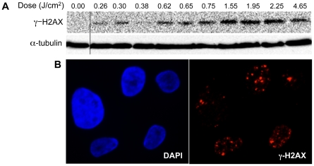
Figure 2. Induction of DNA damage by DBD plasma.
(A) MCF10A cells were treated with the indicated dose of plasma. After one-hour incubation, lysates were prepared and resolved by SDS-PAGE and representative immunoblots with antibody to γ-H2AX (top) or α-tubulin (bottom) are shown. (B) Indirect immunofluorescence was performed utilizing an antibody to γ-H2AX one hour after treatment of MCF10A cells with 1.55 J/cm2 DBD plasma.

