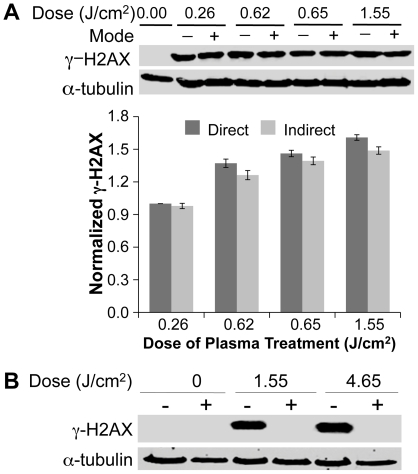
Figure 3. Effects of DBD plasma are mediated by neutral species and not UV generated by plasma in gas phase.
(A) Cells were subjected to plasma as described earlier (direct, D) or a grounded mesh that filters charged particles was placed between the electrode and the medium (indirect, I). Representative immunoblots with γ-H2AX (upper panel) or α-tubulin (lower panel) are shown. The graphs below the immunoblots show quantification from three independent experiments using the Odyssey Infrared Imaging System (LI-COR Biosciences, Lincoln, NE, USA). The γ-H2AX signal was normalized to the amount of α-tubulin and data are expressed relative to lowest dose that was set at 1.0. (B) UV produced in DBD plasma does not induce the observed DNA damage. Cells overlaid with 100 µl of medium were treated with plasma at 1.55 J/cm2 and 4.65 J/cm2 with (+) and without (−) placing magnesium fluoride (MgF2) glass over the cells during treatment. MgF2 glass blocks all plasma species except UV from reaching the surface of the medium covering the cells during treatment. Representative immunoblot with γ-H2AX (upper panel) or α-tubulin (lower panel) is shown.

