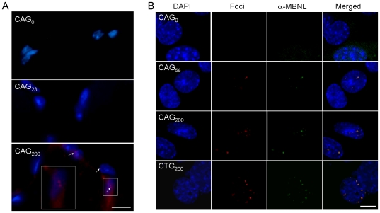
Figure 8. Fluorescence in situ hybridization detection of nuclear foci formation.
(A) Frozen muscle sections from CAG0, CAG23 and CAG200 mice were hybridized with a Cy3-(CTG)13 probe (red). The nuclei were counterstained with DAPI (blue). Merged images of red and blue signals are shown. Nuclear foci (arrows) are only detected in CAG200 sections. (B) C2C12 cells transfected with pEGFP-CAG0/58/200 and pEGFP-CTG200 were hybridized with Cy3-(CTG)13 and Cy3-(CAG)13 probes, respectively. Distribution of endogenous MBNL proteins was visualized by immunostaining with an anti-MBNL antibody and a FITC-conjugated secondary antibody (green). Merged images showing superimposition of red and green signals (yellow) demonstrate that MBNL proteins are colocalized with expanded CAG and CUG repeats. Scale bars, 10 µm.

