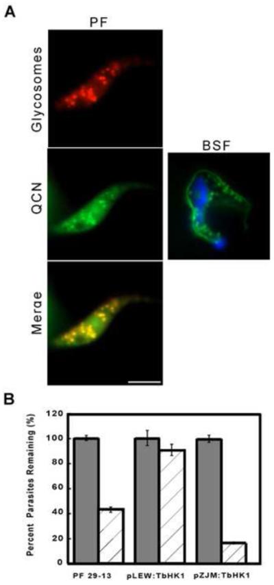
Fig. 3.

Localization and genetic manipulation studies to explore the biological consequences of QCN on T. brucei. BSF and PF parasites incubated with QCN (100 μM, 15 and 10 min for PF and BSF, respectively) were stained with DAPI and imaged. Glycosome localization in live PF cells was visualized by mCherry emission. Fluorescence of the QCN does not bleed into the mCherry spectra and was captured using a 488 nm filter set. Scale bar = 10 μm. (B) RNAi of TbHK1 enhances QCN toxicity, while over-expression of TbHK1 tempers it. Parental PF trypanosomes (29-13) or parasites transformed with pLEW111(2T7):TbHK1 or pZJM:TbHK1 were induced with tetracycline (1 μg/ml) for 24 hours to either express or silence TbHK1, QCN (50 μM, dashed columns) added, and cell viability scored after an additional 24 hour incubation. All assays were performed in triplicate, with cell numbers normalized to untreated controls. The p-values for untreated and treated parental 29-13 and pZJM:TbHK1 bearing cells were statistically significant (p < 0.05 in both cases), indicating that the differences between untreated and treated cells were significant, while the value for pLEW111(2T7):TbHK1 harboring cells incubated with or without QCN was not (p > 0.05).
