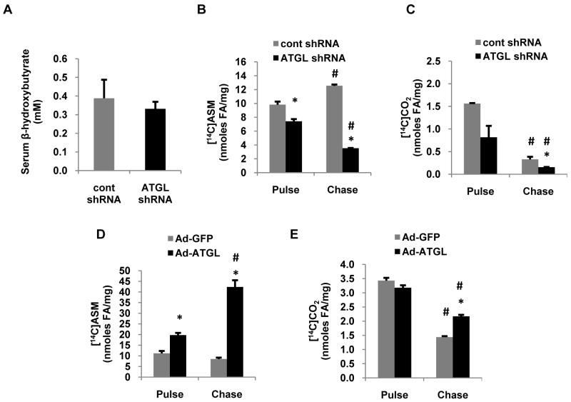
Figure 5. Hepatic ATGL promotes fatty acid oxidation.
(A) Serum β-hydroxybutyrate was measured with a colorimetric enzymatic kit in serum samples collected from chow-fed mice 7 d after infection with control or ATGL shRNA adenovirus and following an overnight fast. (B-E) Primary hepatocytes isolated from chow-fed mice were transduced with control or ATGL shRNA for 66 h and Ad-GFP or Ad-ATGL for 24 h, at which time pulse (1.5 h) and chase (6 h) experiments were performed with 500 μM [1-14C]oleate. Media from hepatocytes were harvested after pulse and chase, and CO2 and ASM were quantified as outlined in the experimental procedures to measure fatty acid oxidation. Data are presented as means ± SEM. *P<0.05 vs control shRNA group. #P<0.05 vs pulse period.

