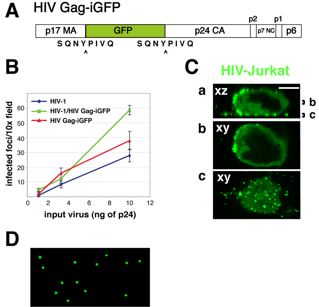
Fig. 1. HIV Gag-iGFP, an infectious, replication competent clone of HIV-1.
(A) Diagram of the Gag-internal, interdomain GFP insertion strategy into HIV Gag. GFP is placed between the MA and CA regions of Gag and flanked by viral protease cleavage sites. Upon maturation, the GFP is cleaved from Gag, but remains associated with the viral particle. (B) HelaT4 Magi cells [24] were infected with HIV Gag-iGFP, parent virus NL4-3, or a mix of the two viruses for 48 hr and then stained for β-galactosidase. (C) Jurkat T cells were transfected with HIV Gag-iGFP as described in the text. After 48 hrs, cells were plated on poly-l-lysine coated coverslips and fixed with 4% paraformaldehyde. Note the accumulation of Gag at the plasma membrane. (D) 293T cells expressing HIV Gag-iGFP following transfection using a calcium phosphate based method. After 48 hrs, supernatants were filtered and spotted onto glass coverslips. Viral particles were fixed and imaged on a confocal microscope using a 63× oil objective and 6× frame averaging.

