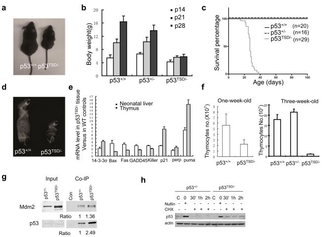
Figure 1.
p53TSD/− mice exhibit segmental progeria and constitutively active p53. (a) Representative image of male p53+/+ and p53TSD/− littermates at 30 days of age. (b) Growth rate of p53+/+, p53+/− and p53TSD/− mice between 14 and 28 days postnatal. Values represent means and s.d. (n = 11). (c) Survival curve of p53+/+-, p53+/−-and p53TSD/−-mice. P < 0.0001 between p53TSD/− mice, compared with p53+/+-and p53+/−-mice. (d) Representative X-ray image showing the spine curvature of 30-day-old p53+/+-and p53TSD/−-littermates. (e) mRNA levels of various p53-target genes in the liver of newborn, and thymus of three-week-old, p53TSD/− mice. The level of mRNA in p53TSD/− tissues was calculated by normalization to wild-type controls. Values represent means ± s.d. (n = 3). (f) The number of thymocytes in mice with the indicated phenotypes at one-week old and at three-weeks old. Values are means ± s.d. (n = 3). (g) The T21D and S23D mutation in p53 partially disrupts the interaction between Mdm2 and p53. To allow normal expression of p53 from the targeted allele, p53TSD/− and p53+/− MEFs were infected with retroviral-Cre to excise the PGK–Neor cassette from the targeted allele. p53 was immunoprecipitated, and the amount of p53 and Mdm2 in the immunoprecipitate was determined by western blotting. Nonspecific antibody (control) was used to indicate the specificity of immunoprecipitation. The ratio of the protein levels of p53 and Mdm2 in the immunoprecipitates from the different mice strains is indicated. (h) Nutlin, which inhibits the interaction between p53 and Mdm2, can stabilize p53TSD and make it more stable than wild-type p53. After deletion of PGK–Neor from the targeted allele, p53TSD/− and p53+/− MEFs were treated with nutlin (25 μM) for 24 h, and then further cultured in medium containing cycloheximide (CHX; 10 μM), but without nutlin. The protein levels of p53 in untreated samples, nutlin-treated samples before cycloheximide treatment (0 h), and samples at the indicated times after cycloheximide treatment, were analysed by western blotting.

