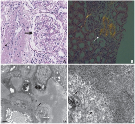
Fig. 5.
A: microscopic findings of renal tissue. PAS-negative eosinophilic material deposition is observed on the wall (thin arrow) of the interlobular artery and glomerular mesangium (thick arrow) (PAS, ×400). B: view of renal tissue under polarizing microscopy. The interlobular artery shows apple-green birefringence (arrow) on Congo-red staining. C: electron microscopic findings of renal tissue showing tangles of fibrils within mesangial area (arrows) (EM, ×12,500). D: high power view of mesangial tangle showing haphazardly arranged fibrils (arrows) (EM, ×30,000).

