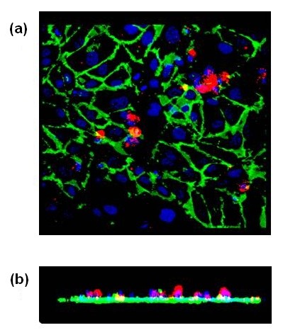
Figure 2.

Image of hMSCs adherent to a confluent monolayer of HUVECs treated with vWF. Confocal image of a planar (a) and Z-axis (b) projections of hMSCs adherent to HUVECs treated with 4 μg/ml vWF for four hours. HUVECs were stained with AF488-conjugated CD31 (green). HMSCs were labeled with PE-conjugated CD90 (red). HMSCs were found on the top of endothelial monolayer within the boundaries of ECs.
