Figure 7.

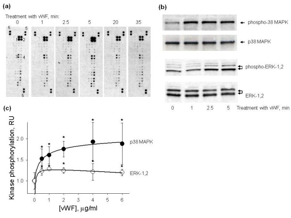
Phosphorylation of protein kinases in HUVECs treated with vWF. Protein phosphorylation in HUVECs treated with vWF was analysed using the human phospho-MAPK array, by Western blot and cell-based ELISAs. (a) Shows the phosphorylation of eighteen protein kinases in HUVECs treated with 4 μg/ml vWF for 0 to 35 minutes assayed using the human phospho-MAPK array. VWF stimulated the phosphorylation of ERK-2 (spots 1), ERK-1 (spots 2), p38α (spots 3) and p38γ (spots 4). Spots labelled with number 5 are the positive controls used for the normalization of the arrays. (b) Shows Western blots of total p38 MAPK and ERK-1,2 and phosphorylated p38 MAPK and ERK-1,2 from lysates of HUVECs treated with 4 μg/ml vWF for 0-5 min. (c) Shows the dose-response curves of p38 MAPK (black circle) and ERK-1,2 (white circle) phosphorylation in HUVECs treated with 0 to 6 μg/ml vWF for four hours measured using the cell-based ELISAs. Data are shown as mean ± SD of four independent measurements. Asterisks mark statistically significant changes in comparison with none treated HUVECs (t-test, P-value <0.05).
