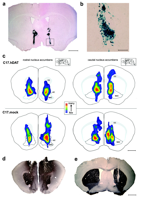
Figure 4.
Histologic analysis of grafted neural stem cells. (a) A representative coronal section from a mouse transplanted with C17.hDAT clone. Section stained for β-galactosidase by using x-gal histochemistry revealed bilateral distribution of surviving grafted cells 10 days after transplantation. (b) Magnified area of the graft shows cells with various morphologies; some extending processes. (c) Summary of the localization of transplanted cells. Relative abundance of x-gal-positive cells in all mice in both groups receiving cell transplants is represented as a colorized xyz isocontour plot (for details, refer to Materials and methods). Cumulative scores from three sections per mouse ranged from few cells (1, blue) to many densely packed cells (5, red). For clarity, details of this method are illustrated in example micrographs in Supplementary Figure S2 in Additional file 3. (d) Coronal section at the level of NAC stained with an antibody against DAT. Although the antibodies used do not distinguish endogenous DAT from that found in the transplanted C17.hDAT cells, the ectopic expression of DAT evident in the cortical region of the transplant track is consistent with the exogenous hDAT. Such ectopic placement of a transplant is not representative of other animals examined in this study. (e) In contrast, a transplant of the C17.mock stem cells into striatum, shown here in a coronal section, is not labeled with the anti-DAT antibody and is clearly visible as a vertical clear track in the right hemisphere, which labeled strongly, revealing the endogenous DAT. CC, corpus callosum; AC, anterior commissure. Scale bars = 1 mm for panels a and c through e; and 200 μm for panel b.

