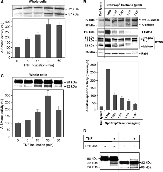
Figure 3.
A-SMase cleavage and activation after TNF stimulation. (A) HeLa cells were stimulated with TNF for indicated times and cell lysates were analysed for endogenous A-SMase by western blotting using anti-A-SMase antibodies that detect the 72-kDa pro-A-SMase and a 57-kDa cleavage product. A-SMase processing is accompanied by an increase of A-SMase activity. Data are from four experiments (+s.e.m.), each performed in triplicates. (B) Cell lysate and OptiPrep fractions from unstimulated HeLa cells were analysed by western blotting using the indicated antibodies and A-SMase assay. A-SMase activity peaked in the lysomal fraction 2. (C) HeLa cells expressing recombinant A-SMase-GFP were stimulated with TNF for indicated times. Western blots were performed using anti-GFP antibodies, detecting pro-A-SMase-GFP at 98 kDa and the cleavage product of A-SMase-EGFP at 82 kDa, which is also accompanied by an increase of A-SMase activity. Data are shown for three experiments (±s.e.m.), each performed in triplicates. (D) Immunoprecipitated pro-A-SMase-EGFP of untreated or TNF-treated cells was deglycosylated by PNGase F treatment and detected by anti-GFP antibody in western blot. Glycosylated pro-A-SMase-EGFP migrates at 98 kDa. The glycosylated cleavage fragment as well as the deglycosylated pro-A-SMase-EGFP migrate at 82 kDa. The deglycosylated cleavage fragment of A-SMase-EGFP migrates at 68 kDa (figure assembled from cropped lanes of the same gel (see primary scans in Supplementary data)).

