Figure 5.
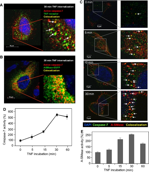
Partial colocalization of active caspase-7 with TNF receptosomes and pro-A-SMase-EGFP as well as with endogenous A-SMase. Merged confocal microscopic images of (A) HeLa cells fluorescence labelled with biotin-TNF/avidin-FITC complexes (green) and anti-cleaved capase-7 monoclonal antibody (Cell signaling) (red) after 30 min of TNF-receptor internalization. (B) HeLa cells labelled with anti-cleaved capase-7 monoclonal antibody (red) and EGFP-tagged A-SMase (green) 20 min after TNF-receptor internalization, (C) HeLa cells labelled with anti-capase-7 antibody (Abcam) (green) and antibodies against endogenous A-SMase at various time points after TNF-receptor internalization. Colocalization of the respective fluorescently labelled molecules is indicated by yellow colour (marked with arrows). (D, E) TNF induction of caspase-7 and A-SMase run in parallel. Pro-A-SMase-EGFP-transfected cells were incubated with TNF for the indicated times. Cell lysates were used for caspase-7 activity assays (D) and A-SMase activity assays (E). Both time courses show a time-dependent increase of activity with the maximum after 30 min of TNF incubation. Data from three experiments (±s.e.m.) are shown.

