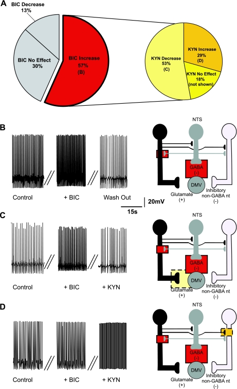
Fig. 2.
Pie charts (A), representative traces (B–D, left), and the proposed neuronal circuits (B–D, right) in neurons in which perfusion with bicuculline (BIC) increased the firing rate of DMV neurons. Perfusion with BIC increased the firing frequency of DMV neurons (N = 35), and perfusion with kynurenic acid (KYN) decreased the firing frequency of the majority of these neurons. Note that the pie chart on the right represents subgroups of neurons in which BIC increased the firing frequency. The increase in firing rate induced by BIC is likely due to antagonism of GABAA receptors located either on the membrane of the DMV neuron or on tonically active glutamatergic neurons that impinge on the DMV neuron. Perfusion with KYN in the presence of BIC decreased the firing rate of DMV neurons (N = 19). This response was likely due to KYN-mediated inhibition of a tonic glutamatergic input onto DMV neurons. Perfusion with KYN in the presence of BIC further increased the firing rate of DMV neurons (N = 10), suggesting the presence of tonic glutamatergic synapses impinging onto non-GABAergic neurons. In all figures, the proposed site of action of bicuculline is indicated by a solid-line red square and the proposed site of action of kynurenic acid is indicated by a dotted-line yellow square.

