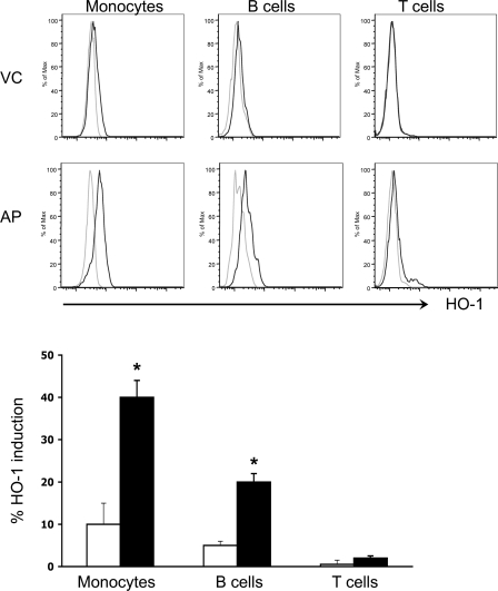
Fig. 4.
PH-mediated HO-1 induction in leukocyte subsets from AP patients and healthy VC. PBMCs from AP patients at the time of admission and from VC donors were isolated by density centrifugation. PBMC were cultured for 24 h with either PH or VE alone. The cells were subsequently stained for leukocyte surface markers followed by staining for intracellular HO-1. The immune-stained cells were then analyzed by FACS. Histograms with light and dark lines are from VE- and PH-treated cells, respectively. Bar graph shows % HO-1 induction as determined from the FACS histograms. Solid and open bars represent results from AP and VC donors, respectively. *P < 0.05.

