Fig. 5.
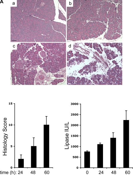
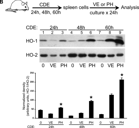
PH-mediated HO-1 induction in mouse splenocytes during experimental pancreatitis. A: FVB/n mice were fed regular chow (a) or CDE for 24 (b), 48 (c), or 60 h (d) to induce mild, moderate, and severe pancreatitis, respectively. Pancreata were isolated and fixed in 10% formalin, and sections were stained with hematoxylin and eosin and scored. Serum was used for determination of lipase levels. Scale bar, 50 μm. B: leukocytes from mouse spleens of the FVB/n mice fed CDE for 24, 48, or 60 h were isolated and cultured for 24 h with either PH or VE alone. Cultured leukocytes were lysed in a homogenization buffer as described in materials and methods, and 10 μg total protein from each sample was separated by SDS-PAGE followed by Western blot analysis to compare HO-1 protein expression levels. Equal protein loading was confirmed by assessing levels of the constitutively-expressed HO-2 isoform. Bar graphs show the density of HO-1 relative to HO-2. *P < 0.05 compared with VE.


