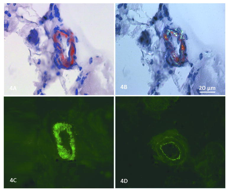
Figure 4.
(A) Congo red stain shows amyloid deposited in the smooth muscle layer of a skeletal muscle arteriole in patient 3. (B) Using polarized light, the green birefringence of amyloid is seen. (C) In a consecutive serial section, dysferlin deposition is seen in this same blood vessel (NCL-Hamlet Novocastra Laboratories Ltd.). (D) In the same section a blood vessel wall lacks dysferlin deposition; only the autofluorescence of the internal elastic membrane (tunica intima) and adventitia (tunica externa) can be seen, but the smooth muscle of the tunica media is devoid of dysferlin.

