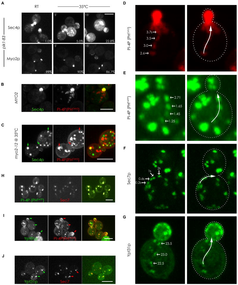
Figure 3. PI4P is present in compartments of the late secretory pathway and is required for their transport.
(A) pik1-83 cells expressing either GFP-Sec4p or Myo2p-GFP were fixed and examined at different times after shifting to 35°C and the percentage of small/medium budded cells with delocalized GFP-Sec4p and polarized Myo2p-3XGFP was determined (percentages in lower right corners). (B) Small budded wild type cell expressing GFP-Sec4p and mCherry-PHOsh2p. (C) myo2-12 cell expressing GFP-Sec4p and mCherry-PHOsh2p after 15-min shift to the restrictive temperature. Arrows indicate examples of colocalization. (D and E) Frames from Supplementary movie 1 (D) and 2 (E) showing vesicular-like structures labeled by the indicated PI4P reporters moving rapidly towards the bud. Times in the movies are indicated. (F) Frames from Supplementary movie 3 showing the directed movement of GFP-Sec7p towards the bud. (G) Frames from Supplementary movie 4 showing the directed movement of the GFP-Ypt31p towards the bud. (H, I, and J) Cells co-expressing a PI4P reporter and different secretory markers showing colocalization (arrows) of PI4P with Sec7p (H), with Ypt31p (I), or between Sec7p and Ypt31p (J). See also Figure S3. Bars represent 5μm.

