Figure 1.
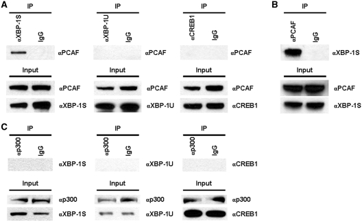
PCAF associates with XBP-1S. (A) 293T cells were transfected with an expression plasmid to ectopically express XBP-1S, XBP-1U and CREB1, respectively. IP was performed using the cell lysates prepared from the transfected cells and the indicated antibodies. Normal IgG (IgG) was used as a negative control. The immunoprecipitated complexes and the protein inputs were analyzed by western blotting. (B) The cell lysates of XBP-1S expressing cells were used for IP with an anti-PCAF antibody. The presence of XBP-1S in the immunoprecipitates was determined by western blotting. (C) Cells were co-transfected with a p300 expression vector and an indicated plasmid (i.e. XBP-1S, XBP-1U, and CREB1 plasmids, respectively). IP was carried out using an anti-p300 antibody followed by western blotting.

