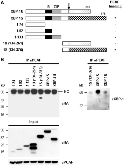
Figure 2.
Domain study of XBP-1. (A) Diagram of XBP-1 truncations. All the constructs were HA-tagged. (B) 293T cells were transfected with the indicated plasmid to express an individual XBP-1 deletion. IP was performed using the anti-PCAF antibody followed by western blotting with anti-HA or anti-XBP-1 antibodies.

