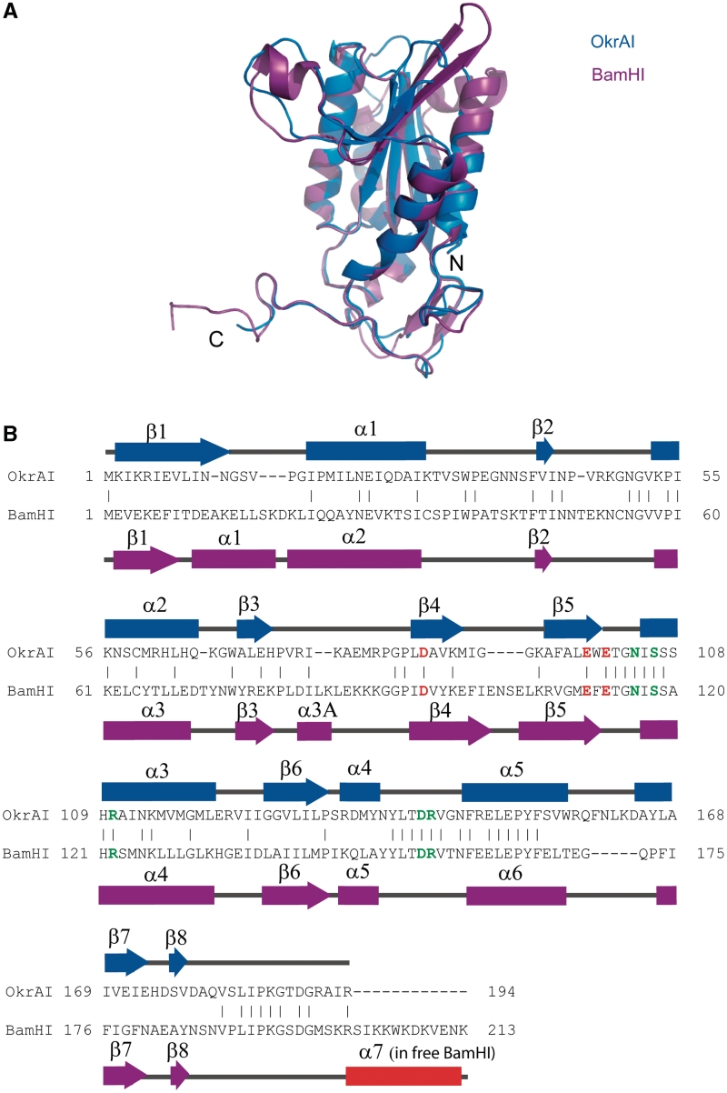
Figure 2.
Comparisons between OkrAI and BamHI subunits. (A) The monomer of OkrAI (blue) superimposed on the monomer of BamHI (magenta). (B) Sequence alignment of OkrAI and BamHI based on the structural alignment performed by DALI (36). The catalytic residues of both enzymes are shown in red and the DNA-binding residues in green. The secondary structural elements are labeled and shown above (OkrAI) and below (BamHI) the alignment, respectively. The BamHI C terminus that is α helical in the apo form and unstructured in the DNA-bound form is highlighted in red. This segment is missing in OkrAI.

