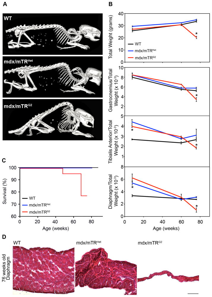
Figure 3. Muscular dystrophy in mdx/mTRG2 mice progresses with age.
(A) Whole body SPECT/CT images. Pronounced skeletal deformity of the spine (kyphosis) is present in 76 week old mdx/mTRG2 mice, compared to age-matched controls.
(B) Animal weight significantly decreased in mdx/mTRG2 mice at 76 weeks of age (top, n≥3, P<0.05). Indicated muscles were harvested from mice at various ages and weighed. Data are represented as the weight of the tissue relative to total body weight, a standard control for telomere shortening (Lee et al., 1998). Results are shown as average±s.e.m (n≥3, P<0.05).
(C) Kaplan-Meyer survival curve. mdx/mTRG2 mice exhibited reduced life-span compared to mdx/mTRHet and WT controls (n≥12).
(D) Hematoxylin/eosin staining of diaphragm muscle from animals at 76 weeks of age showed dramatic atrophy of the tissue in mdx/mTRG2 animals compared to controls (Scale Bar=120μm). See also Fig. S3.

