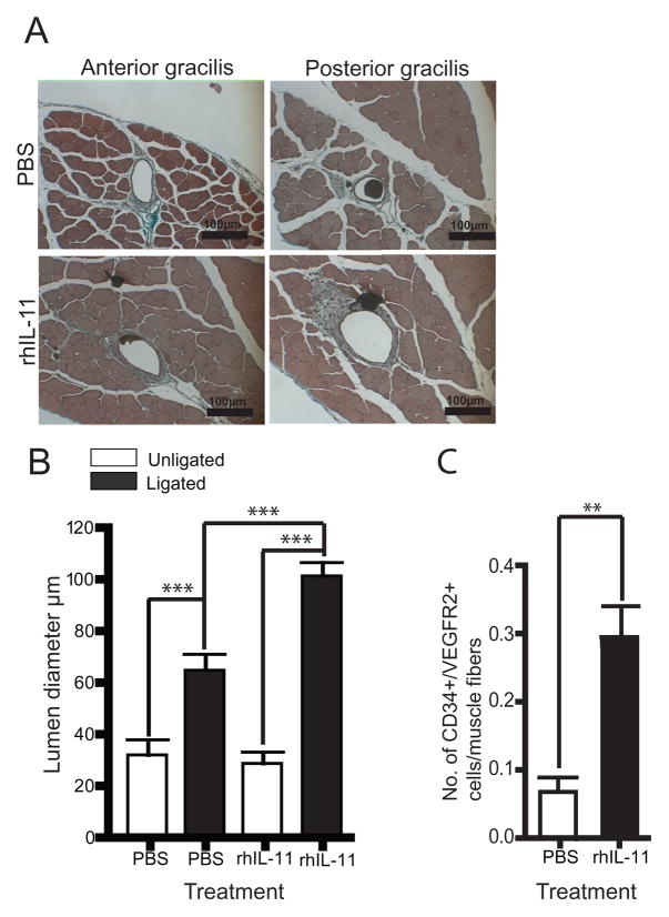
Figure 5. Mice treated with rhIL-11 have increased collateral vessel luminal diameter and influx of perivascular CD34+/VEGFR2+ cells.
A. Cyano-Masson-Elastin staining of anterior and posterior gracilis muscle showing increased collateral vessel luminal diameter in rhIL-11-treated mice 21 days after femoral artery ligation. B. Graph showing that rhIL-11-treated mice have a 3-fold increase in luminal diameter of adductor collateral vessel compared to PBS control. Each bar depicts mean ± SEM of 9 mice. Unpaired student t-test ***=P< 0.0001 (20 × magnification). C. Anterior and posterior gracilis muscle were stained for influx of perivascular CD34+/VEGFR2+ cells. Mice treated with rhIL-11 have 4.4-fold increase in perivascular CD34+/VEGFR2+ cells 8 days after femoral artery ligation. Each bar depicts mean ± SEM of 8 mice. Unpaired student T-test, two tailed **= P< 0.01.

