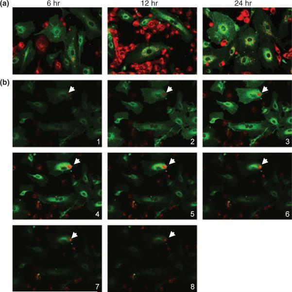
Fig. 1.
Phagocytosis of apoptotic trophoblast cell by endothelial cell in vitro. PKH26-labeled H8 cells (red) were overlaid on 70% confluent PKH67-labeled human endometrial endothelial cells (HEECs) (green) in EBM-2 supplemented with 10% fetal bovine serum and 800 ng/mL puromycin and observed under confocal microscope at different time points (magnification: 400×). (a) Coculture of HEECs with apoptotic H8 cells for 6 hr results in engulfment of trophoblast fragment by endothelial cell. More phagocytosed particles appear in the lysosome around the nucleus after 12 and 24 hr of coculture. (b) Serial z-stack images (from top to bottom, 1 μm apart) of HEECs coculture with apoptotic H8 cells. Note trophoblast-derived particle locates inside the endothelial cells (arrows).

