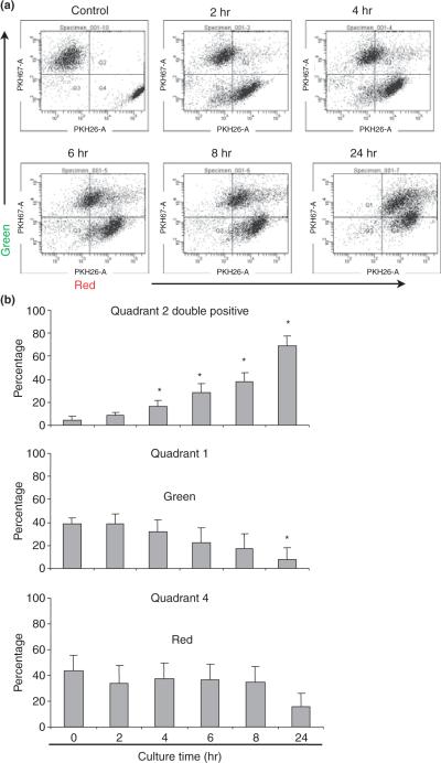
Fig. 2.
Flowcytometric analyze of trophoblast cell and endothelial cells co culture in vitro. PKH26-labeled H8 cells (red) were overlaid on 70% confluent PKH67-labeled human endometrial endothelial cells (HEECs) (green) in EBM-2 supplemented with 10% fetal bovine serum and 800 ng/mL puromycin. Cells were harvested and analyzed using flowcytometry after 2, 4, 6, 8, and 24 hr of coculture. Negative control was prepared by mixing HEECs with H8 cells just before flowcytometry. (a) Control and 2, 4, 6, 8, and 24 hr of HEECs and H8 cells coculture. Quadrant 1 (upper left): green fluorescent HEECs only; Quadrant 2 (upper right): double fluorescent HEECs phagocytosed apoptotic H8 cells; Quadrant 4 (lower left): red fluorescent H8 cells only. (b) Bar chart shows the average percentage of HEECs phagocytosed apoptotic H8 cells (Quadrant 2), HEECs (Quadrant 1) and H8 cells (Quadrant 4) at different time point of coculture. Data are presented as mean ± S.D.; *P < 0.05 relative to control.

