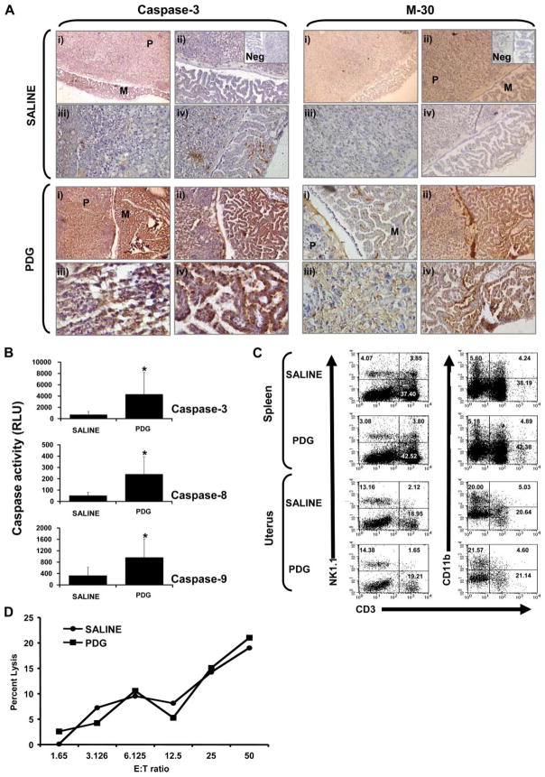
FIGURE 1.
Effect of PDG on placental apoptosis and immune cell recruitment in vivo. C57BL/6 pregnant mice were treated i.p. with either saline or PDG (25 μg/mouse) in 100 μl on embryonic day 6.5. On embryonic day 12, animals were sacrificed and the spleens and fetoplacental units were harvested. A, Paraffin-embedded placental tissue sections were immunostained for active caspase 3 and cell death using the apoptosis marker M-30. For each treatment (saline or PDG) and marker (caspase 3 and M-30), panels are labeled as: i, Placenta labyrinth (P) and fetal membranes (M) (original magnification, ×10); ii, placenta labyrinth and fetal membranes (original magnification, ×20); iii, placental labyrinth (original magnification, ×40); and iv, fetal membranes (original magnification, ×20–40). Insets show the Ab-negative controls (Neg). B, Placental tissue lysates were evaluated for activation of the caspase pathway using the Caspase-Glo assay. Bar graphs show caspase 3, caspase 8, and caspase 9 activities as RLU. Value of p < 0.05 relative to the saline controls. C, The proportion of uterine and splenic NK cells were evaluated using flow cytometry by first gating on the CD45+ cells and then by analyzing CD3, NK1.1, and CD11b expression. As shown on the scatter graphs, the proportion of uterine and splenic NK cells (NK1.1+CD3−) and myeloid cells (CD11b+CD3−) were similar between the mice treated with saline and those treated with PDG. C, Uterine NK cells were isolated and their cytotoxicity was tested against YAC-1 cells. D, Bar graph, Similar levels of uterine NK cell cytotoxicity from mice treated with either saline or PDG.

