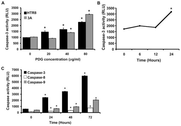
FIGURE 2.
Effect of PDG on trophoblast apoptosis in vitro. A, First trimester trophoblast cells (HTR8 and 3A) were incubated with PDG (0–80 μg/ml) for 48 h, after which cell lysates were prepared and caspase 3 activity was determined using the Caspase-Glo assay. Bar graphs show caspase activity in RLU. PDG significantly increased trophoblast cell caspase 3 activity in a dose-dependent manner. *, p < 0.001 relative to the untreated control (0). B, First trimester trophoblast cells (HTR8) were incubated with PDG (80 μg/ml) and, after 0, 6, 12, and 24 h, cell lysates were prepared and caspase 3 activity was evaluated. C, First trimester trophoblast cells (HTR8) were incubated with PDG (80 μg/ml). After 0, 24, 48, and 72 h, cell lysates were prepared and caspase 3, caspase 8, and caspase 9 activity was evaluated. PDG significantly increased trophoblast cell caspase activity in a time- dependent manner. *, p < 0.001 relative to time 0.

