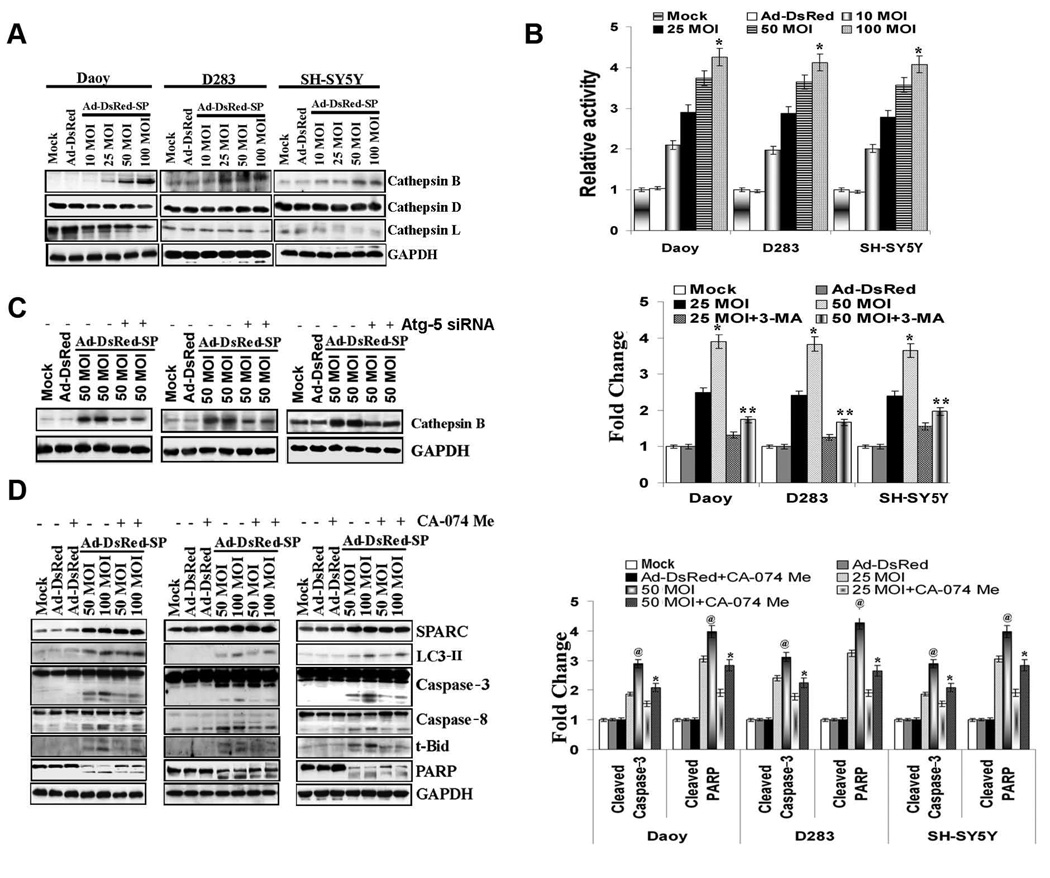
Figure 4. Cathepsin B mediates SPARC-induced apoptosis in PNET cells.
PNET cells were infected with mock, 100 MOI of Ad-DsRed, and the indicated MOI of Ad-DsRed-SP for 48 h. (A) Cathepsin (B, D and L) levels were assessed by Western blot analysis in cell lysates. (B) PNET cells were infected as mentioned above and Cathepsin B activity was measured. (C) PNET cells were infected with mock, 100 MOI of Ad-DsRed, and the indicated MOI of Ad-DsRed-SP and treated with autophagy inhibitor, 3-MA. Levels of cathepsin B were determined by Western blot analysis. (D) PNET cells were infected with mock, 100 MOI of Ad-DsRed, and the indicated MOI of Ad-DsRed-SP and treated with 100 µM cathepsin B inhibitor CA-074 Me. Cells were harvested and levels of cleaved caspases 8 and 3, PARP and t-Bid were assessed by western blot analysis. Results are representative of three independent experiments. GAPDH served as a loading control. (Columns, mean of three experiments; bars, SD)

