Figure 3.
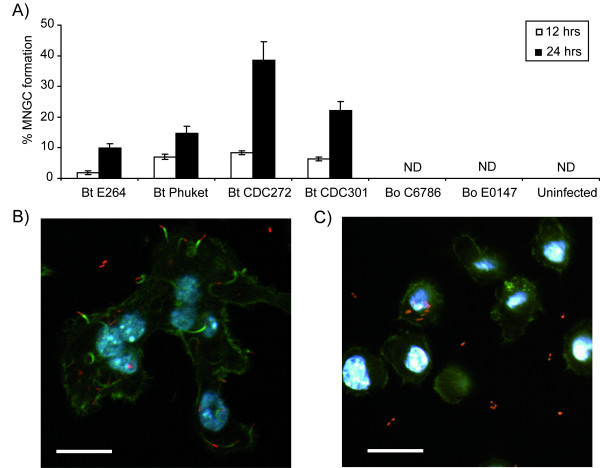
MNGC formation and intracellular behaviour of Burkholderia strains in macrophages. J774A.1 macrophages were infected with Burkholderia strains at an MOI of 10 as already described. (A) Multinucleated giant cell (MNGC) formation was assessed at 12 hrs and 24 hrs post infection. Cells were stained with Giemsa and the percentage of MNGCs was calculated relative to normal macrophages per field of view. MNGCs were defined as cells containing 3 or more nuclei. The error bars represent the standard error of the mean derived from at least 10 fields of view. ND = not detected. (B-C) Representative confocal micrographs of cells at 8 hrs post infection with B. thailandensis strain E264 (B) and B. oklahomensis strain C6786 (C). In both panels, bacteria appear red due to expression of RFP from the modified broad-host-range vector pBHR4-groS-RFP. Filamentous actin was stained green with FITC-phalloidin conjugate and nuclei were stained with DAPI. Scale bars represent 20 μm.

