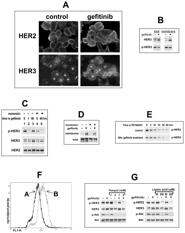
Figure 3. Mechanism of HER3 reactivation following extended HER TKI treatment.
(A) SKBr3 cells were treated with 5 μM gefitinib or control for 48h and stained with anti-HER2 or anti-HER3 antibodies for immunofluorescence microscopy. (B) In addition, cell surface proteins in control or 48 hour pre-treated cells were biotinylated, precipitated, and immunoblotted as indicated. (C) Control or gefitinib pre-treated (5 μM, 48h) SKBr3 cells were treated with 20 μM monensin for the final 6h and analyzed by western blotting. (D) SkBr3 cells were treated with gefitinib for 48 hrs and with 20uM monensin for the final 6 hours. Membrane and total HER3 was immunoblotted from cell surface proteome pulldowns (above) or total lysates (below). (E) The dephosphorylation rate of p-HER3 following 48hrs of gefitinib treatment was determined immediately after initiation of fully inactivating concentrations of the irreversible HER TK inhibitor PD168393 (2 μM). (F) Reactive oxygen species were quantified in control (A) or gefitinib pre-treated (B) (5 μM, 48h)SKBr3 cells as described in methods. (G) SKBr3 cells were treated with 5 uM gefitinib or in combination with the indicated concentrations of the anti-oxidants Tempol or α-lipoic acid for 48 hours.

