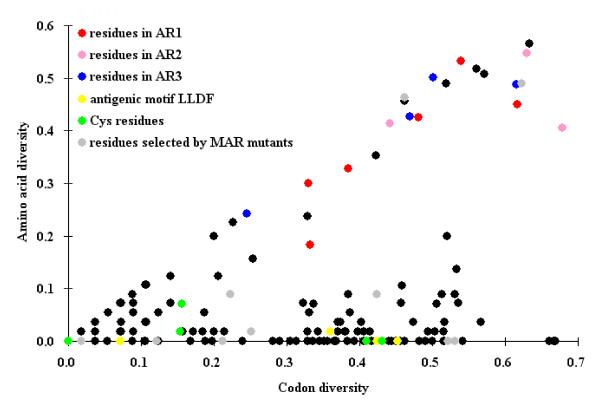
Figure 6.
Analysis of codon and amino acid diversity of residues within the antigenic units of glycoprotein E2. Codon and amino acid diversity was quantified using a modified Simpson's index [41]. Antigenic residues identified in this study are colored according to the antigenic regions (AR) where they occur. Residues of the antigenic motif of 771LLFD774 [25], the six conserved cysteine residues and the antigenic residues identified by mAb-resistant (MAR) mutants analysis [22], are marked in yellow, green and grey, respectively. The other residues are shown in black.

